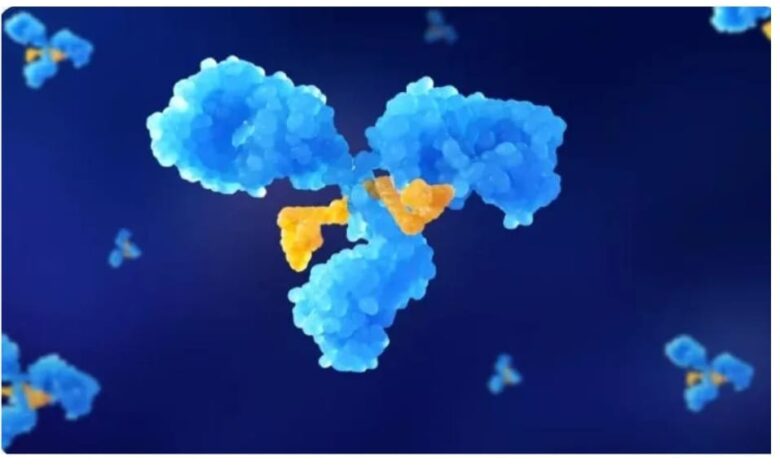
Antibody‑drug conjugates (ADCs) link a monoclonal antibody to a potent cytotoxic payload through a specialized linker. After injection, this complex must survive in circulation, find tumor cells, enter them, and then release the drug inside specific compartments. Each step affects safety, efficacy, and dosing schedules in the clinic. The antibody guides the payload to tumor‑associated antigens, while the linker controls when and where the drug becomes active. An accurate view of this journey helps researchers design better ADCs and helps clinicians understand variable patient responses, off‑target effects, and resistance. The following sections track this path from the bloodstream to the lysosome.
Systemic Circulation and Distribution
Entry into Bloodstream
After intravenous injection, the ADC enters the systemic circulation almost immediately as an intact macromolecule. The antibody component dominates its pharmacokinetic behavior, so the conjugate distributes primarily within the vascular and interstitial spaces rather than inside cells. Blood flow carries the ADC through the heart, lungs, liver, kidneys, and peripheral tissues. The large molecular size limits rapid diffusion across tight endothelial barriers, but fenestrated or leaky vessels permit some tissue penetration. Fc regions of the antibody interact with the neonatal Fc receptor (FcRn), which recycles the ADC and extends its half-life. This recycling process helps maintain therapeutic plasma concentrations long enough for the ADC to encounter tumor tissue, bind its antigen, and initiate downstream internalization and adc payload.
Plasma Stability and Protection
Once in circulation, the ADC must remain stable to prevent premature payload release, which can cause systemic toxicity and reduce tumor exposure. The linker chemistry plays a central role here. Non‑cleavable linkers resist enzymatic and chemical degradation in plasma, while cleavable linkers are designed to respond mainly to tumor‑associated conditions such as low pH or specific proteases. Albumin and other serum proteins may weakly interact with the ADC surface, but the antibody structure typically preserves its binding specificity. Enzymes, reducing agents, and mechanical shear all challenge conjugate integrity over time. Robust design minimizes deconjugation and aggregation, keeping the payload firmly attached until the ADC reaches antigen‑expressing cells and undergoes endocytosis and lysosomal processing.
Tumor Targeting and Accumulation
Antigen Recognition and Binding
As the ADC circulates, the antibody component seeks cells that express the target antigen at the surface. Tumor cells often overexpress these antigens compared with normal tissues, creating a concentration gradient that favors selective binding. When the antibody encounters its antigen, noncovalent interactions form a tight complex with high affinity and specificity. This binding step anchors the ADC on the tumor cell membrane and initiates clustering of antigen–ADC complexes. The degree of antigen expression, accessibility on the cell surface, and internalization rate all influence how much ADC accumulates in the tumor. Heterogeneous antigen distribution within solid tumors can lead to variable uptake, but strong and sustained binding is still a key driver of targeted payload delivery.
Enhanced Permeability and Retention Effect
Solid tumors often exhibit abnormal, leaky vasculature and impaired lymphatic drainage. This combination underlies the enhanced permeability and retention (EPR) effect, which allows large molecules such as ADCs to accumulate more in tumor tissue than in most healthy organs. Gaps between endothelial cells permit macromolecules to extravasate into the tumor interstitium. Because lymphatic clearance is poor, these agents tend to remain trapped in the tumor microenvironment for extended periods. The EPR effect does not act alone; it works together with antigen‑mediated binding to increase local ADC concentrations. However, its magnitude varies widely among tumor types and individual patients, so ADC design and dosing must also account for inconsistent vascular permeability and interstitial pressure within tumors.

Cellular Uptake and Processing
Endocytosis into Target Cells
After antigen binding, the cell internalizes the ADC–antigen complex through receptor‑mediated endocytosis. Clathrin‑coated pits often mediate this process, but other endocytic pathways can also contribute depending on the antigen. The plasma membrane invaginates, pinches off, and forms an early endosome containing the ADC. The rate of internalization depends on antigen biology, epitope selection, and antibody properties. Efficient internalization increases payload delivery per binding event, while slow internalization can limit cytotoxic impact. Some ADCs exploit antigens that rapidly recycle between the surface and intracellular compartments, which allows repeated uptake cycles. Once inside, the endosomal environment begins to acidify, setting the stage for further trafficking toward lysosomes, where many linkers are designed to undergo controlled cleavage.
Intracellular Trafficking to Lysosomes
Following endocytosis, early endosomes mature into late endosomes and then fuse with lysosomes, which contain hydrolytic enzymes and an acidic pH. Many ADC linkers are engineered to respond to this environment. Protease‑cleavable linkers are cut by lysosomal enzymes such as cathepsins, while acid‑labile linkers respond to the low pH. Non‑cleavable linkers require complete antibody degradation to liberate the active payload‑amino acid complex. Once released, the cytotoxic drug can diffuse into the cytosol and reach its molecular target, such as tubulin or DNA. Some payloads display a bystander effect, crossing cell membranes to affect neighboring tumor cells. The efficiency of lysosomal trafficking and processing thus directly shapes both potency and selectivity of the ADC therapy.
Conclusion
The journey of an ADC payload from injection to intracellular release involves coordinated steps in circulation, tumor targeting, and cellular processing. Antibody structure and FcRn interactions govern systemic exposure, while linker stability preserves the conjugate in plasma. Antigen recognition, EPR‑driven accumulation, and efficient endocytosis then concentrate ADCs in tumor cells. Finally, lysosomal trafficking and linker cleavage determine how much active drug reaches its intracellular targets. Each design choice—antibody, linker, and payload—modulates these stages, influencing therapeutic index and resistance patterns. Understanding this pathway guides rational ADC optimization and supports safer, more effective targeted cancer treatments.